


How do bacteria divide?
Oeiras, 17.08.2015
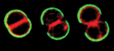
The precision of the new microscopy techniques used in the study now published in Nature Communications show how the currently accepted model for the division of the pathogenic bacteria Staphylococcus aureus needs rethinking. The study involved researchers from the Lab of Bacterial Cell Biology and the Lab of Bacterial Cell Surfaces and Pathogenesis.
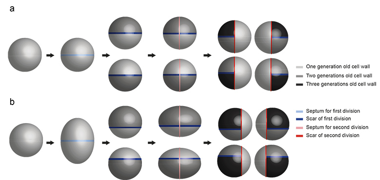
Staph bacteria are spherical. In order for these bacteria to colonize their host, each sphere must give rise to two new spheres, something that occurs every 30-60 minutes. The division takes place in a precise sequence of three perpendicular planes and so far, researchers supposed that the plane for each division was determined by the position of the scars from previous divisions. But the premises for this hypothesis may not be as accurate as they thought. A closer look to the division process revealed that staph bacteria elongate before dividing. By analyzing the bacterial cell wall, researchers also determined that 60% of coating of each sphere originates from the mother sphere. This contradicts what you would expect from a simple equatorial division (50%) and places the scars in a position incompatible with the currently accepted division model. The new spheres are then able to grow in all directions, stretching the old wall and synthesizing new one.
Monteiro et al., Nature Communications
Researchers were able to determine how long it takes between the division of a sphere in two hemispheres and the actual popping of two new spheres. The process is incredibly fast: 2 milliseconds! A time frame that seems incompatible with the need for the activity of enzymes that cleave the bacterial surface and therefore suggests that this is essentially a mechanical process.
“It’s interesting how we were wrong before” says Mariana Gomes de Pinho who coordinated the study “it’s true that conventional microscopy tools did not allow such precise measurements but, if one looks carefully to previous scanning electron microscopy photos, the scars are not really positioned in the equator of the bacteria”. Current super-resolution microscopes (a technique worth the Chemistry Nobel Prize in 2014) show Staph bacteria with an incredible definition and were crucial to acquire the data that must now be taken into account when conceiving a new model to explain how Staph bacteria divide.
Original Article
Nature Communications (2015) DOI: 10.1038/ncomms9055
Cell shape dynamics during the staphylococcal cell cycle
João M. Monteiro*,1, Pedro B. Fernandes*,1, Filipa Vaz2, Ana R. Pereira1, Andreia C. Tavares1, Maria T. Ferreira1, Pedro M. Pereira1, Helena Veiga1, Erkin Kuru3,4, Michael VanNieuwenhze3, Yves V. Brun4, Sérgio R. Filipe2 and Mariana G. Pinho1
1 - Laboratory of Bacterial Cell Biology (ITQB); 2 - Laboratory of Bacterial Cell Surfaces and Pathogenesis (ITQB); 3 - Department of Chemistry, Indiana University; 4 - Department of Biology, Indiana University
* These authors contributed equally to this work
Monteiro et al., Nature Communications


