In vitro Skin development
In vitro human skin models are increasingly gaining attention for their importance in basic research and clinical applications. Recently, this need was amplified due to EU regulations which encourage the replacement and reduction of animal models (EU Directive 2010/63/EU) and ban testing cosmetic products in animals (EU Cosmetic Directive 76/768/EEC, REACH regulation 1907/2006). The use of three dimensional (3D) skin tissue to perform the screening of new drug candidates, physiological evaliation of the tissue and evaluation of therapeutics treatments of dissease phenotypes represents a valuable tool that contribute to the biomedical research without the need of animal testing.
The BMD group is growing in vitro skin since 2016, obtaining reproducible epidermis models and complete epidermis-dermis models with high quality, including co-culture with melanocites and the use of biopolymeric scaffolds developed at the Chemistry Department of the Faculdade de Ciências e Tecnologia Universidade NOVA de Lisboa.
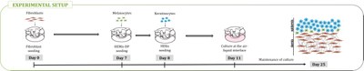
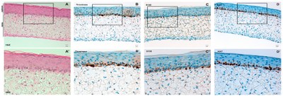
Reconstructed 3D full humanized skin, experimental set up and Reconstructed human dermis and epidermis with haematoxylin-eosin staining (A and A’). Stratification and differentiation of keratinocytes leading to formation of stratum corneum. In the model, the melanocytes are aligned in basal layer and are identified with tyrosinase and S100 proteins by immunohistochemistry (B, B’, C and C’). The viability of the model is demonstrated by the proliferation marker Ki67 (D and D’).
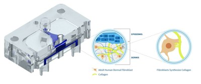
The development of a full controlled microfabricated device for full 3D skin development is under construction. A newly designed prototype has been construted and tested, filled with the dermal scaffold (collagen and fibroblast cells) and seeded with the keratinocytes. Perfusion of media for cell growing and human plasma will allow the control of the skin characteristics, even inducing disease phenotypes.
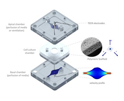
Schematic layout of the microfabricated 3D-skin-in-a-chip.
Keywords: epidermis; reconstructed skin, 3D skin, keratinocytes, organ-in-a-chip


