


Under pressure: how do cells die?
Oeiras, 16th August 2025
Inside cells, the endoplasmic reticulum (ER) works like a factory, producing proteins and folding them into their final shapes, that are essential for their function. But this process may go awry, leading to the accumulation of misfolded, non-functional proteins in the ER and causing stress on cells – ER stress. At first, cells try to adapt by producing machinery to increase its protein folding capacity. However, when ER stress is prolonged and severe, cells may end up programming their own death, so that organisms can survive. The mechanisms that regulate cell death induced by ER stress remain poorly characterized. Learning more about these mechanisms is crucial to develop treatments for diseases where this kind of cell death is relevant, as for example diabetes, where the cells that produce insulin in the pancreas are sensitive to intense ER stress.
Previously, scientists knew that a molecular sensor called Ire1 detects stress in the ER and activates a key molecule, Xbp1, which turns specific genes on or off by binding to DNA. In the new study, a team of researchers from ITQB NOVA and collaborators from other institutions provides an explanation for the long-standing conundrum of how cell death is regulated by Xbp1.
In the new study, published in Nature Communications, the researchers described that when the active form of Xbp1 was expressed in the eyes of fruit flies they ended up with smaller, atrophic eyes, a sign of cell death. By performing a genetic screen, they found out that this trait was reversed when a protein called Fbxo42 was not functional, suggesting that it might somehow interfere with Xbp1’s modus operandi. To discover of how this could be, they went looking for relevant molecules that interact with Fbxo42, and identified Ataxin-2 as a key target. Fbxo42 brings Ataxin-2 to a protein complex that links a small protein called Ubiquitin to Ataxin-2, which is then targeted for degradation by the proteasome, a machinery in cells that degrades proteins that are no longer necessary.
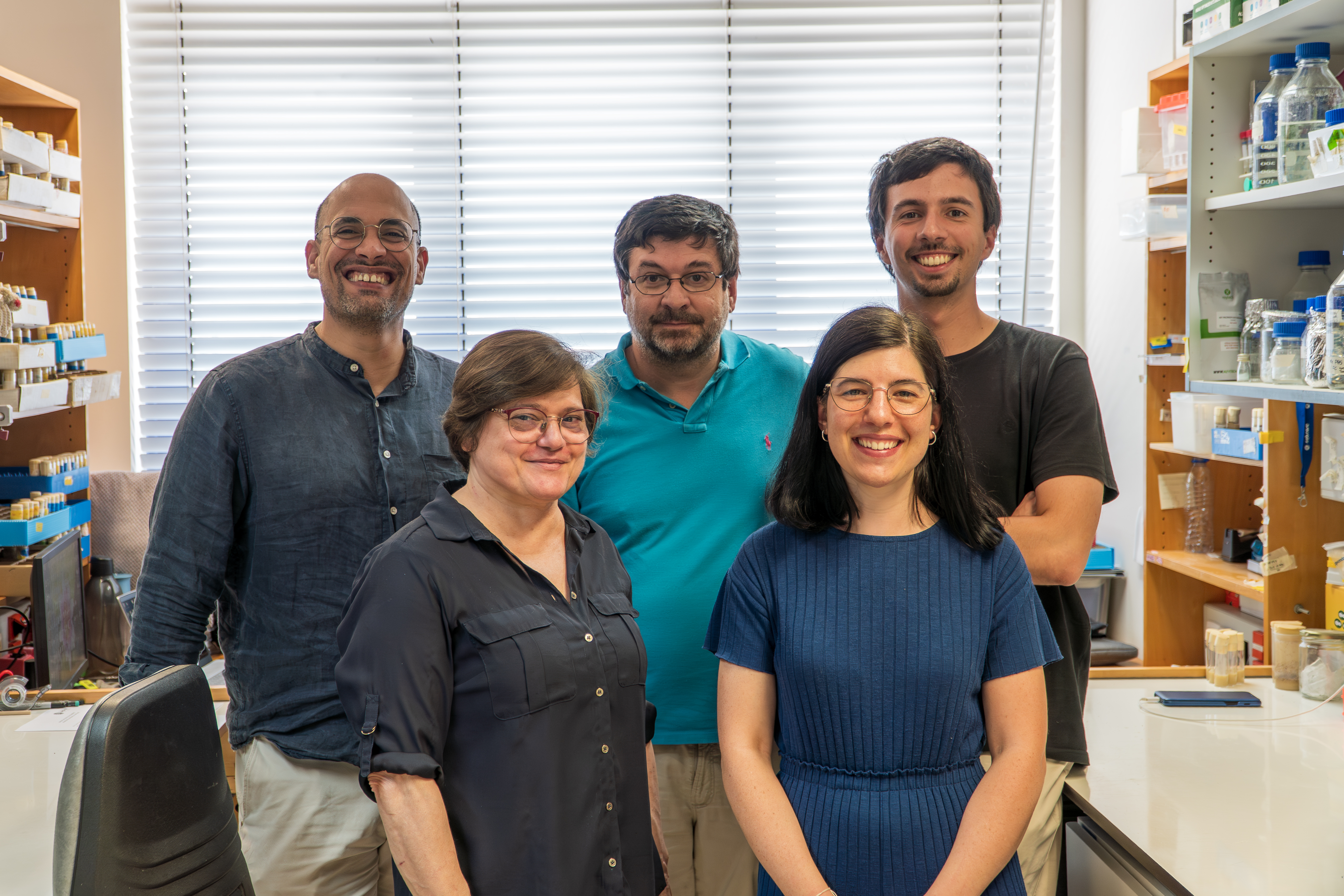
ITQB NOVA Researchers that are part of this study’s team: from left to right, top to bottom: Tiago Cordeiro, Pedro Domingos, Miguel Trigo, Fátima Cairrão, Cristiana Santos.
With this data, the researchers could build a better picture of what is going on inside cells undergoing high levels of ER stress: “In early, pro-survival stages, Ire1 cuts Xbp1’s messenger RNA (mRNA), the instructions the cell uses to make the Xbp1 protein, into its active form, which is then bound by Ataxin-2 and stored in temporary structures inside cells called stress granules. Here, Xbp1 mRNA bound to Ataxin-2 is protected from degradation but it is not translated into Xbp1 protein, preventing cell death in the initial phases, when cells are trying to recover from ER stress”, explains Cristiana Santos, former PhD student at ITQB NOVA and first author of the paper. “In later stages, Fbxo42 is recruited to stress granules, promoting the degradation of Ataxin-2 and releasing Xbp1 mRNA to be translated into Xbp1 protein. The release of this large storage of Xbp1 leads to a terminal wave of signaling inducing the expression of death receptors and committing cells to death”, she adds.
This is the first evidence for a mechanism leading to the degradation of Ataxin-2 by proteasome and the disassembly of stress granules. “This may also have important translational impacts in human diseases, such as diabetes and cancer, where this type of cell death plays a key pathological role, but even more so in neurodegenerative disorders where accumulation of pathological forms of Ataxin-2 causes ataxia and amyotrophic lateral sclerosis. "Next, we have to investigate whether Fbxo42 also promotes the degradation of Ataxin-2 in human cells”, explains Pedro Domingos, senior author of the study and leader of the Cell Signaling in Drosophila lab, at ITQB NOVA.
The study involved two other ITQB NOVA labs: the Dynamic Structural Biology Lab, led by Tiago Cordeiro, and the Single Molecule Microbiology lab, led by Zach Hensel. The study was developed in collaboration with other researchers from the NOVA Medical School, the University of Zurich, Switzerland, the University of the Basque Country, Spain and Queen’s University Belfast, UK.
Original paper:
Nature Communications | https://www.nature.com/articles/s41467-025-62417-2
Fbxo42 promotes the degradation of Ataxin-2 granules to trigger terminal Xbp1 signaling
Cristiana C. Santos, Nadine Schweizer, Fátima Cairrão, Juanma Ramirez, Nerea Osinalde, Ming Yang, Catarina J. Gaspar, Vanya I. Rasheva, Miguel L. Trigo, Zach Hensel, Colin Adrain, Tiago N. Cordeiro, Franka Voigt, Paulo A. Gameiro, Ugo Mayor & Pedro M. Domingos.


